

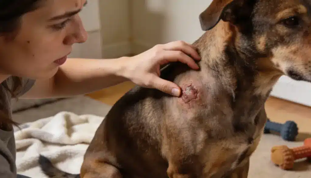
Finding an unfamiliar lump on your dog is unsettling, especially when it changes size, turns red, or looks irritated.
It’s natural to want an answer right away, but here’s the most important thing to know: you can’t tell what a lump is by looking at it.
A mast cell tumor can look almost identical to a harmless bump. Appearance alone never confirms the diagnosis. The only reliable way to know what you’re dealing with is a simple veterinary sample of the cells.
To know more about what mast cell tumors are, the signs to watch for, how vets diagnose and classify them, what a “stage 1” diagnosis actually means, the treatment options available, and what shapes your dog’s long-term outlook, find your answers below.
What is a Mast Cell Tumor?
A mast cell tumor forms when these cells begin growing uncontrollably. A mast cell tumor forms when these cells begin growing uncontrollably.
The American College of Veterinary Surgeons notes in its mast cell tumor guidance that these tumors are the most common type of skin tumor diagnosed in dogs.
They’re diagnosed most often in middle-aged and older dogs, though any dog can develop one. Certain breeds carry a higher risk, including Boxers, Boston Terriers, Pugs, Labrador Retrievers, Golden Retrievers, Rhodesian Ridgebacks, Weimaraners, and some bulldog-type breeds.
That said, breed is only a risk factor. It can neither confirm nor rule out a mast cell tumor on its own.
Symptoms of a Mast Cell Tumor in Dogs
Symptoms vary between dogs. Some develop only a skin lump, while others experience digestive problems or a serious reaction caused by chemicals released from the tumor.
- A raised, flat, soft, or firm skin lump
- Redness or swelling around the affected area
- Hair loss, irritation, or an open surface over the lump
- A mass that suddenly changes size
- Scratching, licking, or rubbing the affected area
- Vomiting or loss of appetite
- Abdominal discomfort
- Black or tar-like stool
- Weakness or rapidly spreading swelling
- Breathing difficulty or collapse
Avoid Handling the Lump: Do not squeeze, massage, or repeatedly touch it. Handling may release chemicals from the mast cells, causing increased redness, swelling, or irritation.
Black or tar-like stool may indicate gastrointestinal bleeding, one of the warning signs of blood loss, and requires prompt veterinary attention. Seek emergency care if your dog develops breathing difficulty, collapses, becomes severely weak, or experiences rapidly spreading swelling.
Stages of Mast Cell Tumors in Dogs
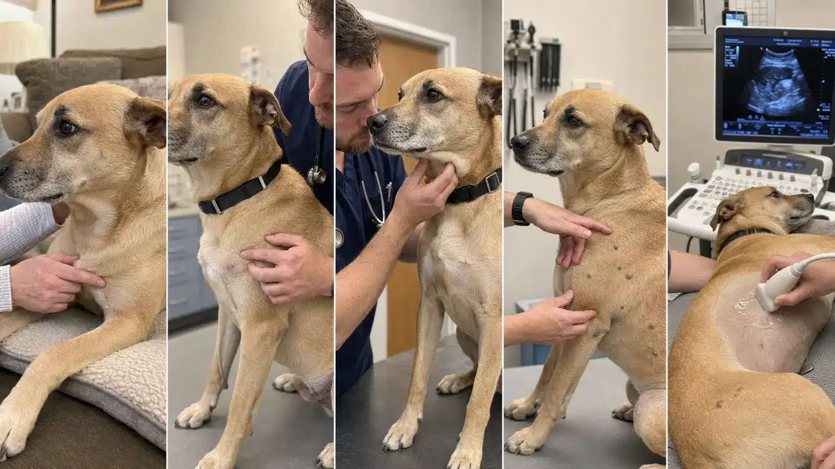
Staging describes how far the cancer has spread, while grading describes how abnormal and aggressive the cells appear under a microscope. The traditional WHO system uses Stages 0 through IV.
Stage 0: Incompletely Removed Tumor
Stage 0 means a single mast cell tumor was removed incompletely, while no involvement was found in the regional lymph node.
Microscopic cancer cells may therefore remain at the surgical site even though spread to nearby nodes has not been identified.
This stage concerns residual local disease after surgery. Tumor grade, margin findings, location, and possible additional treatment still influence what the veterinarian recommends next.
Stage I: One Localized Skin Tumor
Stage I means one mast cell tumor is confined to the skin, with no identified spread to the regional lymph node. It does not mean Patnaik grade I or guarantee a low-grade, slow-moving tumor.
A localized Stage I tumor can still be high-grade under the microscope. Stage describes where the disease is, while grade indicates how abnormal and aggressive the tumor cells appear.
Stages I through III may also receive an a or b substage. Substage a means the dog has no systemic illness, while substage b indicates whole-body symptoms such as vomiting, appetite loss, black stool, or weakness.
Stage II: Regional Lymph Node Involvement
Stage II means one skin mast cell tumor is present and cancer cells have reached the regional draining lymph node.
The primary tumor remains solitary, but lymph node involvement confirms spread beyond the original site.
This finding may affect treatment and prognosis, so the veterinarian considers the involved node, tumor grade, surgical margins, and other test results before recommending a treatment plan.
Stage III: Multiple or Infiltrating Tumors
Stage III includes multiple mast cell tumors in the skin or one large, infiltrating tumor, with or without regional lymph node involvement.
It represents more extensive local or regional disease than Stages I and II, but does not define microscopic aggressiveness.
The veterinarian considers tumor number, size, invasiveness, lymph node findings, grade, location, and whether complete surgical removal appears possible when planning treatment.
Stage IV: Distant Metastasis
Stage IV means distant metastasis is present, potentially involving the spleen, liver, blood, bone marrow, or another site beyond the primary tumor and regional lymph node.
It represents the most widely spread category in the traditional system. Imaging, tissue or needle samples, blood tests, tumor grade, symptoms, treatment response, and the dog’s overall health all help the veterinarian plan appropriate care.
Note: A higher stage generally means the disease is more extensive, but it cannot predict the outcome alone. Grade, tumor location, surgical margins, lymph node findings, cell-division rate, and treatment response also affect prognosis.
Checking Whether It Has Spread
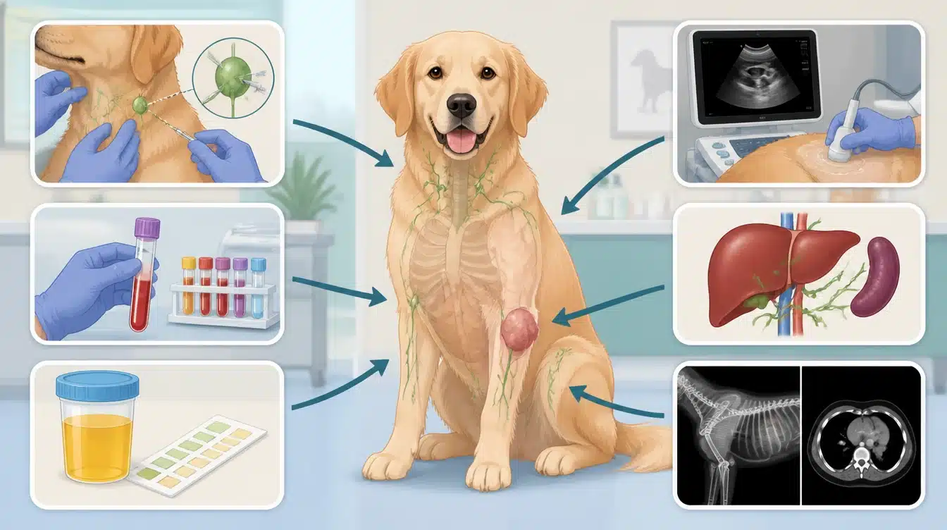
Staging tests are chosen based on the tumor’s grade, location, size, your dog’s symptoms, and whether the results would actually change the plan. Depending on the case, they may include:
- Sampling the regional or mapped sentinel lymph node
- Blood and urine tests
- Abdominal ultrasound
- Selected liver or spleen samples
- Additional imaging if distant disease or another condition is suspected
One point often surprises pet parents: a normal-sized lymph node isn’t necessarily tumor-free. That’s why a vet may recommend sampling the node that drains the tumor area even when it looks and feels perfectly normal.
How Vets Diagnose a Mast Cell Tumor
Diagnosis starts with a physical exam and a sample of the lump’s cells. If mast cells turn up, further testing helps determine the tumor’s grade, how far it extends locally, and whether it has spread.
Step 1: Fine-Needle Aspiration (FNA)
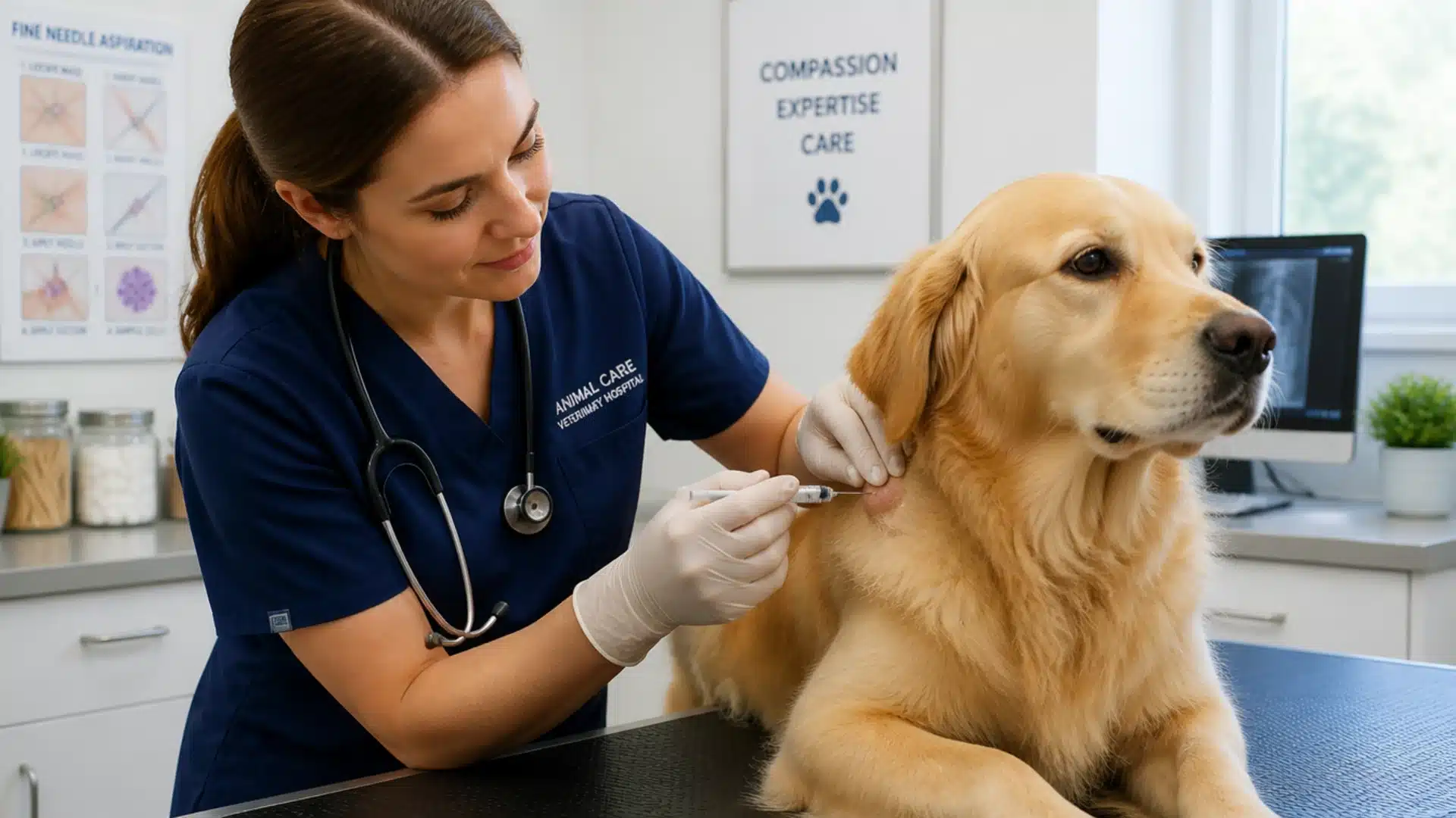
- Purpose: Fine-needle aspiration is usually the first test used when a veterinarian suspects a mast cell tumor.
- Process: A thin needle collects a small sample of cells from the lump, which is placed on a slide and examined under a microscope.
- Result: The procedure is quick, usually needs no sedation, causes minimal discomfort, and can identify mast cells before surgery.
Step 2: Biopsy and Histopathology
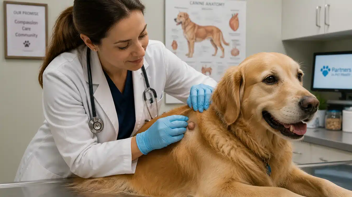
- Purpose: Biopsy and histopathology examine a larger tissue sample or the entire mass after surgical removal.
- Process: A veterinary pathologist studies cell appearance, tumor grade, mitotic activity, depth of invasion, and the condition of the surgical margins.
- Result: These findings help predict tumor behavior, show whether cancer cells may remain, and guide decisions about further surgery, radiation, or medication.
A Note on Grading Systems: You may see two grading systems on the same report, and they are not interchangeable:
- The Kiupel system sorts skin mast cell tumors into low-grade or high-grade.
- The older Patnaik system uses grades 1, 2, and 3.
A report may list both, but a Patnaik grade can’t be swapped one-for-one with a Kiupel result. If you’re unsure how they relate in your dog’s case, ask your vet to walk you through it.
Treatment Measures for Your Dog
There’s no one-size-fits-all treatment. The right plan depends on the tumor’s location, size, grade, stage, margins, lymph-node findings, and your dog’s general health. Some dogs need surgery alone; others need a combination of approaches.
| Treatment | When it may be considered | Main goal |
|---|---|---|
| Surgery | A localized tumor that can be removed | Remove the primary tumor |
| Second surgery | Incomplete margins, with enough tissue to remove more | Clear remaining local cells |
| Radiation therapy | Incomplete removal or a hard-to-reach location | Improve local control |
| Chemotherapy | High-grade tumor or disease that has spread | Treat cancer beyond the original site |
| Targeted medicine | Selected recurrent, non-removable, or advanced tumors | Block signals driving tumor growth |
| Local injection | Eligible non-metastatic tumors, in approved settings | Destroy the tumor locally |
| Supportive medicine | Histamine-related inflammation or stomach upset | Manage tumor-related side effects |
The pathology report will describe the margins as complete, narrow, or incomplete. If cells may have been left behind and there’s enough tissue to work with, your vet may discuss a second operation. When complete surgery isn’t possible, radiation can improve local control.
Chemotherapy is often advised for high-grade tumors or documented spread, while tyrosine kinase inhibitors, studied through the National Institutes of Health’s comparative oncology research, target specific growth signals.
An approved intratumoral injection is suitable for certain non-metastatic tumors. Your vet may also prescribe antihistamines or stomach-protective medications to manage the effects of mast cell degranulation.
Medication Note: Never give human antihistamines, pain relievers, stomach medicines, or supplements without veterinary approval. And don’t stop or change a prescribed cancer treatment unless your vet directs you to.
Yes. They should be merged because life expectancy and follow-up care both explain what happens after treatment. Separate subheadings make the section feel fragmented.
Prognosis and Life After Treatment
Prognosis cannot be judged from clinical stage alone. A stage 1 tumor may have a favorable outlook when it is low-grade and completely removed, while a localized high-grade tumor may require additional treatment despite being assigned the same stage.
Your veterinarian considers several factors together:
- Kiupel or Patnaik grade
- Clinical stage
- Tumor size and location
- Mitotic count
- Surgical margin status
- Lymph-node involvement
- Spread to internal organs
- Recurrence
- Response to treatment
Published survival estimates apply only to dogs with similar tumors and treatment plans. Your veterinarian can interpret those figures using the pathology report, staging results, surgical outcome, and treatment response.
After treatment, regular rechecks help monitor healing, recurrence, and new growths. Between appointments, check your dog’s skin monthly and photograph or measure anything unusual.
Watch the surgical site for swelling, redness, discharge, or reopening, as these changes may indicate a healing problem or infection.
Have every new lump sampled promptly. It may be a separate mast cell tumor rather than a recurrence, and testing is the only way to determine the difference.
Final Thoughts
Mast cell tumors can behave in very different ways, and worth repeating, their appearance says nothing about how serious they are.
A fine-needle aspiration can identify mast cells, tissue pathology provides the grade and margins needed to plan treatment, and staging checks whether the disease has moved beyond the original site.
For some dogs, surgery is enough. Others need additional care. Either way, regular rechecks and monthly skin checks stay important long after treatment ends.
The single most useful thing you can do is simple: have any new, swollen, irritated, or changing lump checked by a vet. Early testing gives you clearer choices and, often, better outcomes.
Frequently Asked Questions
How Quickly Can a Mast Cell Tumor Spread?
It varies widely. Some low-grade tumors stay put for months or years, while high-grade tumors can grow and spread faster. Since appearance can’t predict this, prompt sampling is far more useful than waiting to see how the lump changes.
Can a Dog Live Normally After Mast Cell Tumor Surgery?
Many dogs return to their usual routines once they’ve healed, depending on the tumor and their overall health. Continued rechecks and prompt testing of any new lumps remain important, because another mast cell tumor can develop later.
What Happens if a Mast Cell Tumor isn’t Removed?
Left untreated, it may enlarge, become irritated or ulcerated, cause pain, release histamine, and potentially spread. Surgery isn’t right for every case, so your vet can discuss other local, systemic, or comfort-focused options.
Can Mast Cell Tumors Come Back After Treatment?
Yes. Cells can regrow at or near the treated site, especially if local control was incomplete, and a dog can also develop an unrelated new tumor elsewhere. Testing is what distinguishes a recurrence from a separate new tumor.