

Finding a lump on a dog can instantly raise questions, especially when it seems to appear out of nowhere.
If you’re searching through pictures of dog tumors and cysts, chances are you’re trying to compare what you’ve found and figure out how concerned you should be.
\While I understand how unsettling that moment can feel, photos are only a starting point and cannot replace a veterinary diagnosis. They can, however, help you recognize common skin growths and spot warning signs that deserve prompt attention.
As you learn more about different types of lumps, their appearance, possible causes, and treatment options, you’ll feel better prepared for your veterinary visit and more confident about the next steps.
Causes of Lumps, Bumps, Tumors, and Cysts in Dogs
Dogs can develop lumps, bumps, tumors, and cysts for many different reasons, and not all of them are a cause for concern.
Normal aging often leads to harmless growths like lipomas and skin tags, especially in senior dogs.
Blocked oil glands can trap sebum beneath the skin, forming sebaceous cysts. Fat accumulation may result in soft, movable lipomas that are typically benign.
Infections from bites, wounds, or bacteria can cause swollen abscesses, while inflammation from allergies or irritation may produce temporary lumps.
In some dogs, genetics increase the likelihood of developing certain skin growths. More serious lumps may develop due to cancerous cell growth, making veterinary evaluation essential for any new, changing, or unusual mass.
Understanding the Different Types of Dog Lumps
Not every lump has the same cause or level of concern. Understanding the main categories makes it easier to compare pictures and know when veterinary attention may be needed.
1. Cysts: Cysts are fluid- or oil-filled sacs that usually develop because of blocked oil glands or hair follicles. They often appear as smooth, round, slow-growing lumps and are generally non-cancerous.
2. Benign Tumors: Benign tumors are non-cancerous growths that remain localized. Examples include lipomas and histiocytomas, which typically grow slowly and rarely spread to other tissues.
3. Malignant Tumors: Malignant tumors contain cancerous cells that can invade nearby tissue and spread elsewhere in the body. Their appearance varies, making early veterinary diagnosis essential.
4. Non-Tumor Skin Growths: Abscesses, skin tags, warts, and inflammatory swellings are not true tumors. They often result from infections, irritation, or minor injuries and may still require veterinary evaluation.
Caution: Never attempt to diagnose, drain, or treat a lump on your dog based solely on visual comparison. Many benign and malignant growths can look similar, and only laboratory testing (such as fine needle aspiration or biopsy) can confirm what a lump truly is.
Pictures of Dog Tumors and Cysts: Common Lumps, Bumps, and Skin Growths
Comparing different skin growths can help identify common characteristics before a visit to a veterinarian. While these descriptions support visual comparison, only proper veterinary testing can confirm the exact cause of any lump or bump.
1. Sebaceous Cyst
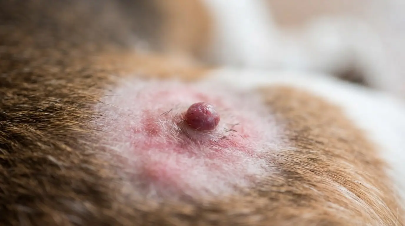
Sebaceous cysts develop when an oil gland becomes blocked, trapping sebum beneath the skin. They usually appear as round, smooth, white, or yellowish lumps and grow slowly over time.
If a cyst ruptures, it may release thick material and become irritated, requiring veterinary evaluation and appropriate treatment.
2. Follicular Cyst
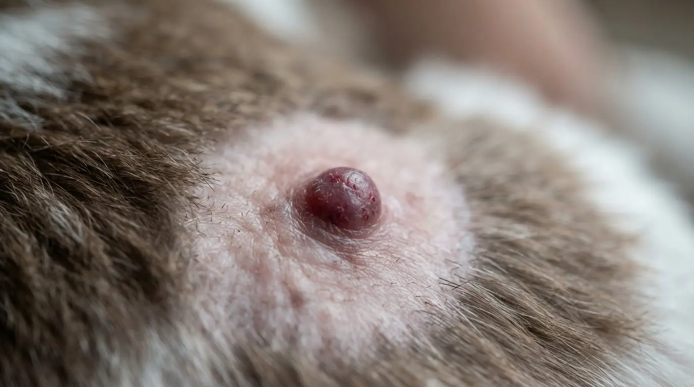
Follicular cysts form within hair follicles after they become blocked or damaged. These cysts typically appear as firm, dome-shaped bumps beneath the skin and may cause localized hair loss.
Although usually harmless, infected or enlarged cysts can become uncomfortable and sometimes require surgical removal by a veterinarian.
3. Epidermal Cyst
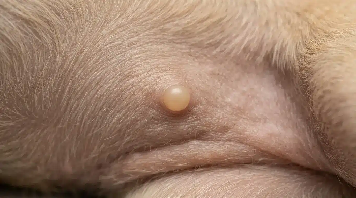
Epidermal cysts occur when skin cells become trapped beneath the surface instead of shedding normally. They often look like small, smooth, flesh-colored lumps with a well-defined shape.
Most grow gradually and remain non-cancerous, though irritation or infection may require professional treatment and monitoring.
4. Apocrine Cyst
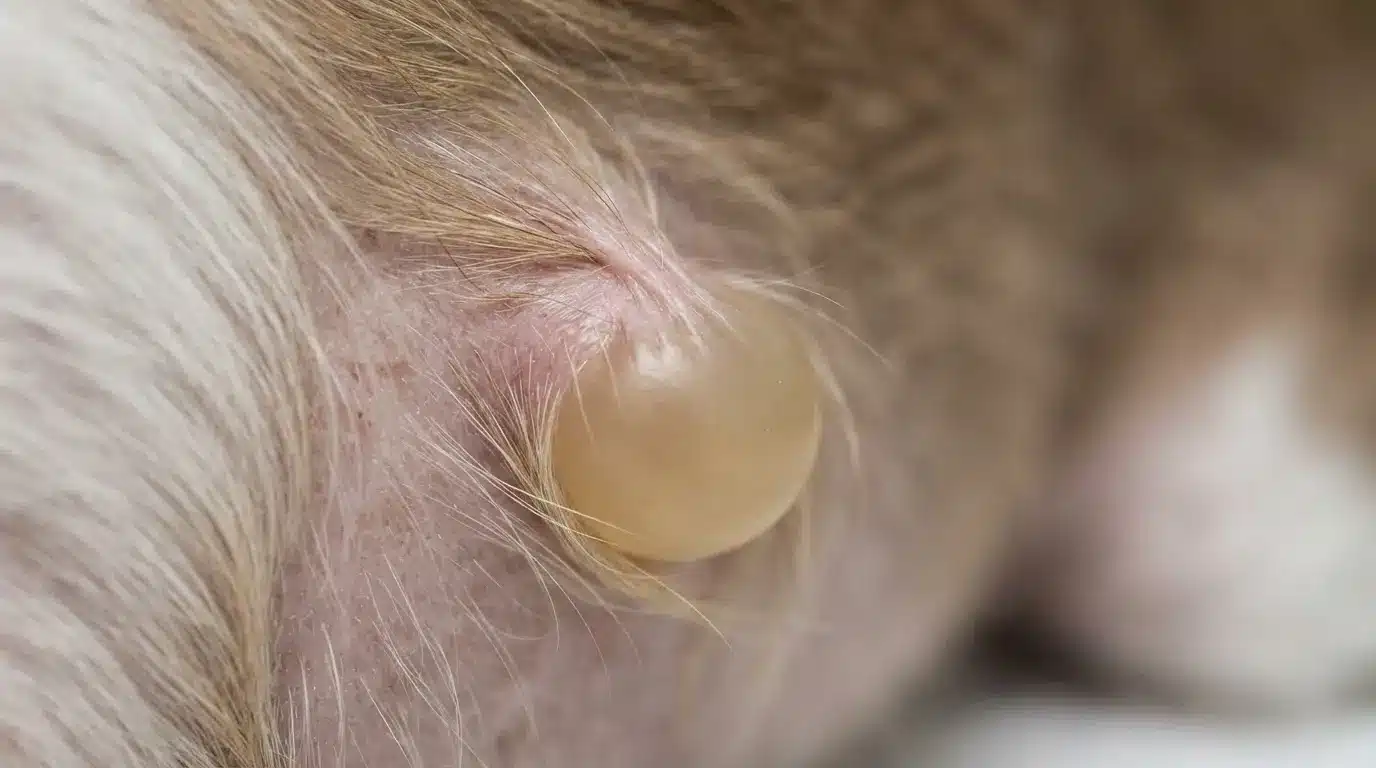
Apocrine cysts originate from the sweat glands located within the skin. These uncommon cysts usually appear as soft, fluid-filled swellings that enlarge slowly.
While they are generally benign, veterinary examination helps distinguish them from other skin masses that may require different forms of treatment.
5. Dermoid Cyst
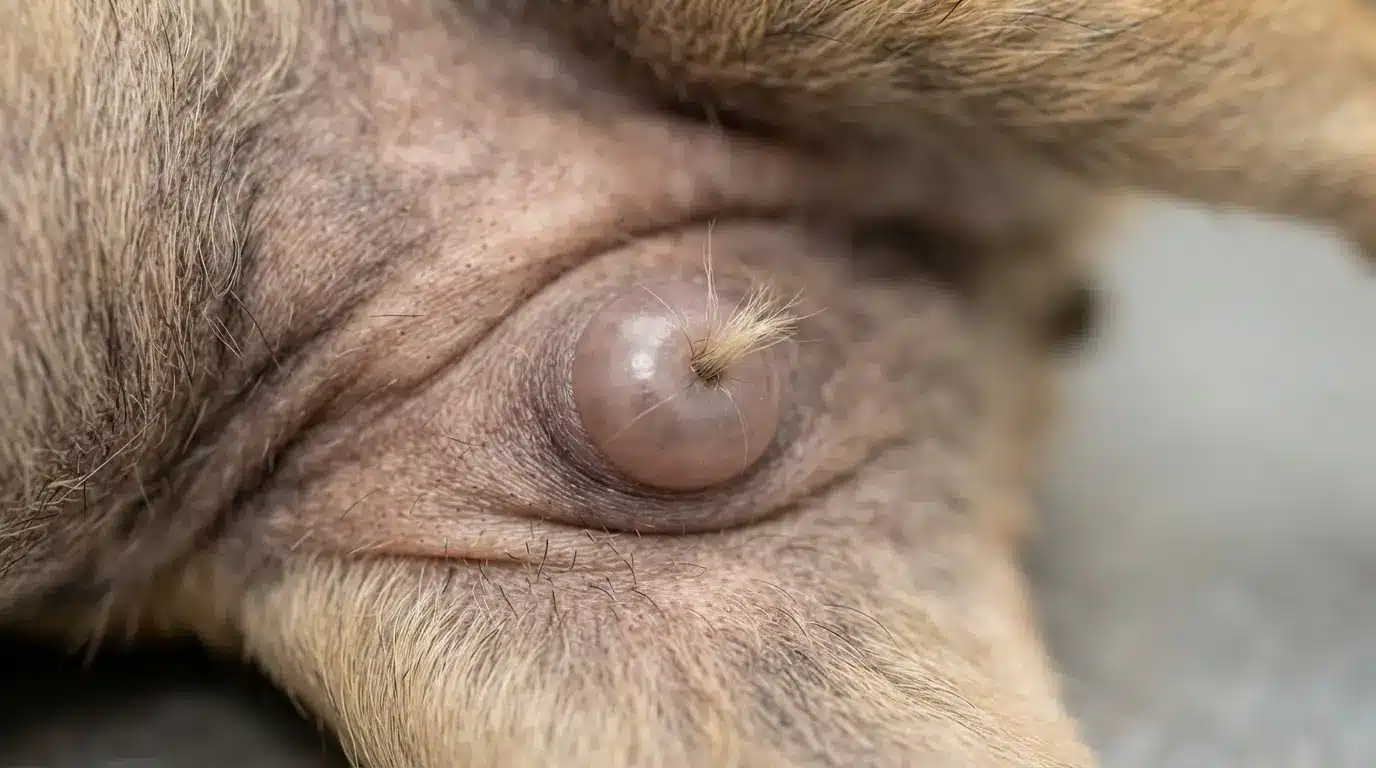
Dermoid cysts are congenital growths, meaning they are present from birth because of abnormal skin development. They often contain hair, skin cells, or other tissues and feel firm beneath the skin.
Surgical removal is commonly recommended if the cyst becomes infected or causes discomfort.
6. Interdigital Cyst
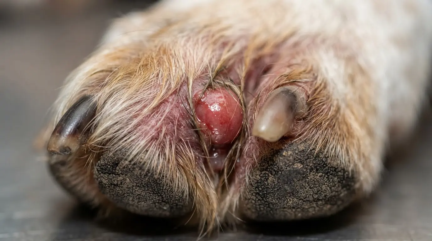
Interdigital cysts develop between a dog’s toes, often because of irritation, infection, allergies, or ingrown hairs. They usually appear as red, swollen, painful bumps that can make walking uncomfortable.
Veterinary treatment helps reduce inflammation, address the underlying cause, and prevent recurring flare-ups.
7. Lipoma (Fatty Tumor)
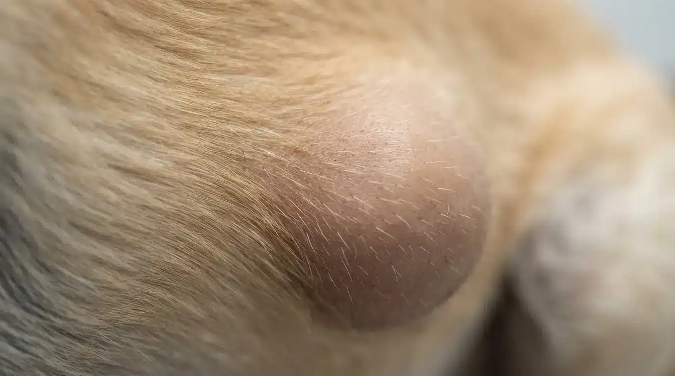
Lipomas are among the most common benign tumors found in older dogs. They usually feel soft, movable, and doughy beneath the skin and often develop on the chest, abdomen, or legs.
Most grow slowly and only require removal if they interfere with movement or become unusually large.
8. Histiocytoma
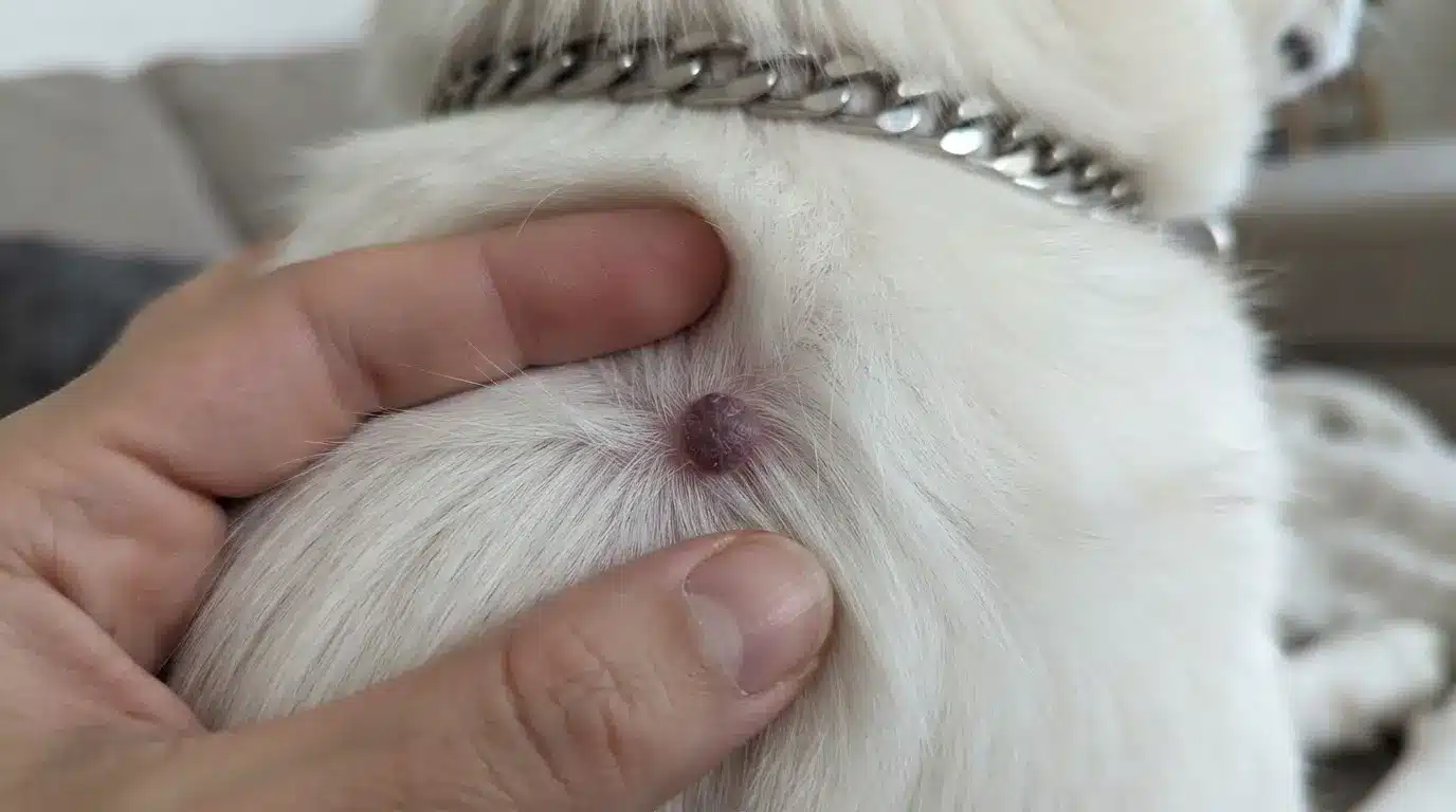
Histiocytomas commonly affect young dogs and appear as small, round, bright red bumps on the face, ears, or legs.
These benign tumors often develop quickly but may disappear on their own within a few months. Veterinary evaluation confirms the diagnosis and rules out more serious growths.
9. Papilloma (Dog Wart)
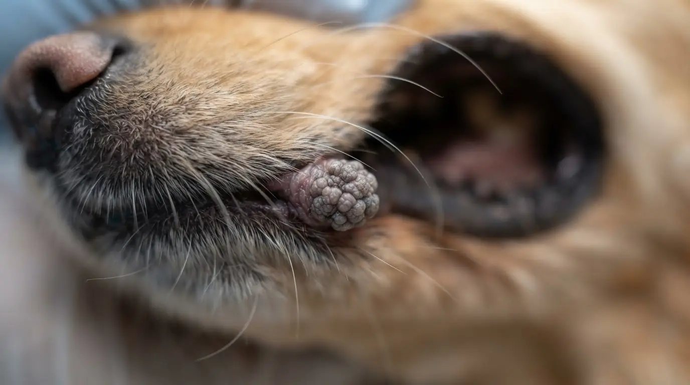
Papillomas are wart-like growths caused by the canine papillomavirus. They usually have a rough, cauliflower-like surface and commonly appear around the mouth, lips, or eyelids.
Most resolve naturally as the immune system responds, although persistent growths may require veterinary removal.
10. Sebaceous Adenoma
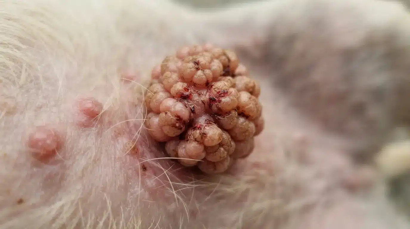
Sebaceous adenomas are benign tumors of the oil glands that frequently develop in senior dogs. They often appear as small, raised, wart-like growths with a rough surface.
Although they rarely become dangerous, removal may be recommended if they bleed, become irritated, or interfere with grooming.
11. Fibroma
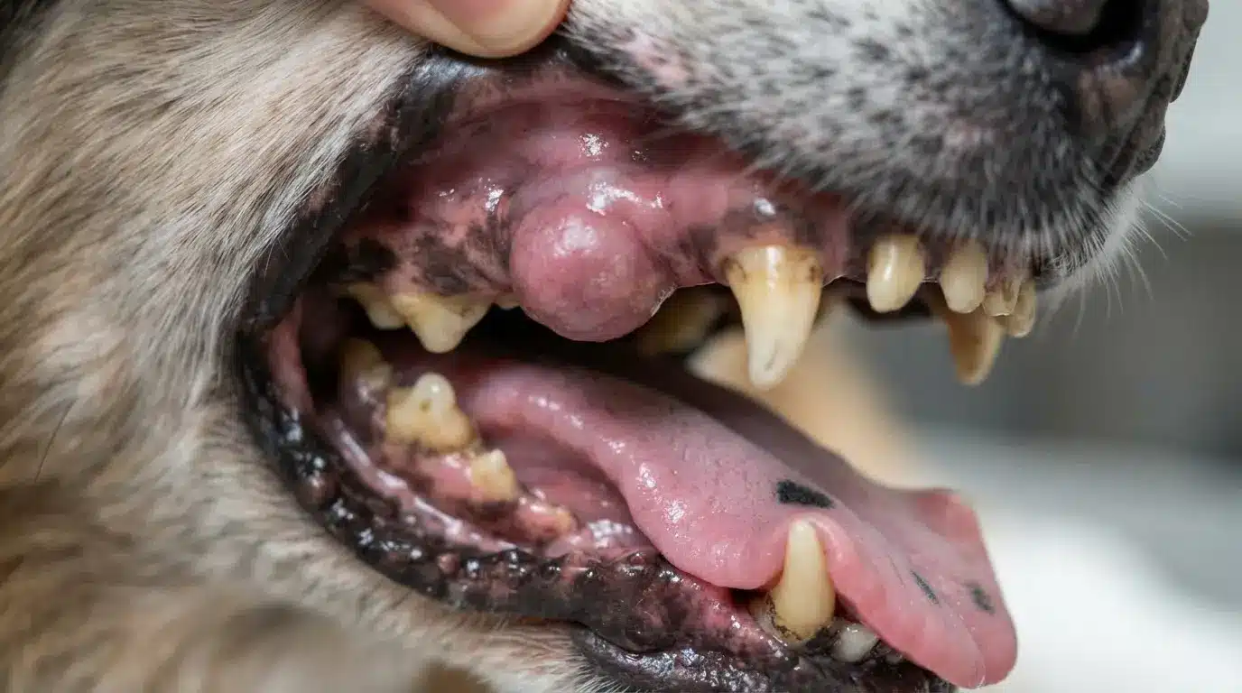
Fibromas are slow-growing benign tumors made of fibrous connective tissue. They usually appear as firm, smooth lumps beneath the skin or on the surface and are commonly found in middle-aged or older dogs.
Surgical removal is considered if the tumor causes irritation or limits normal movement.
12. Perianal Adenoma
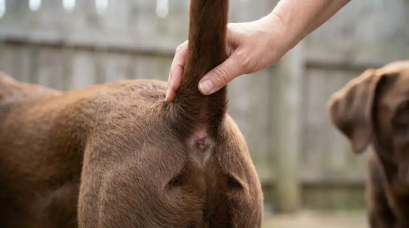
Perianal adenomas develop from the glands surrounding the anus and are most common in older, unneutered male dogs.
These tumors usually appear as small, pink, rounded masses near the rectum. Treatment often involves surgical removal, along with addressing hormonal factors to reduce recurrence.
13. Mast Cell Tumor
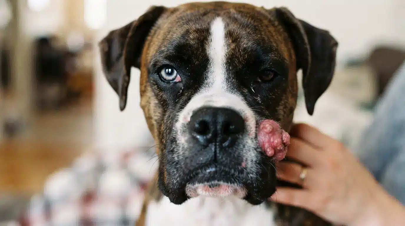
Mast cell tumors are among the most common cancerous skin tumors in dogs. Their appearance varies greatly, ranging from small, hairless bumps to swollen or ulcerated masses.
Because they can closely resemble harmless lumps, immediate veterinary evaluation and biopsy are strongly recommended.
14. Soft Tissue Sarcoma
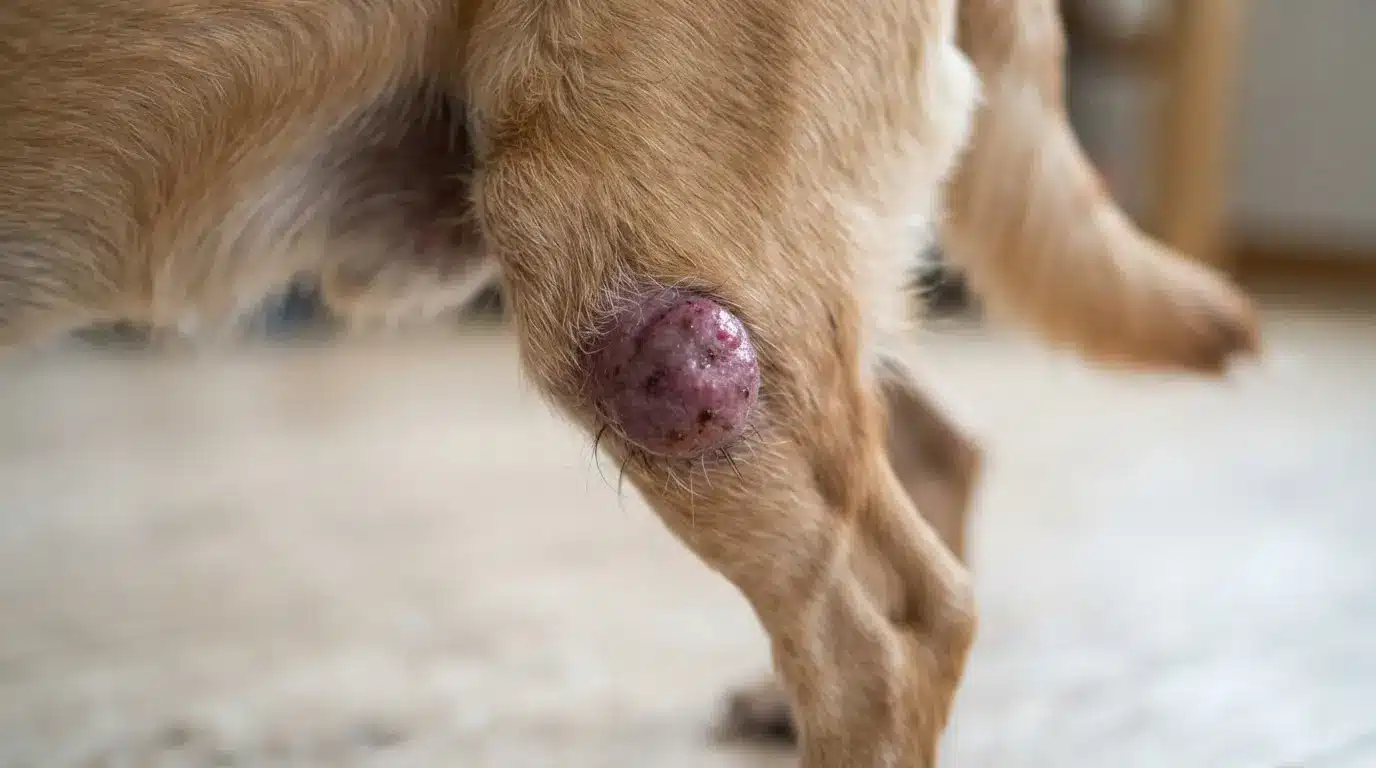
Soft tissue sarcomas develop from connective tissues, including muscles, fat, and nerves. They typically feel firm, fixed beneath the skin, and grow gradually over time.
Although they spread slowly, they often invade nearby tissue, making early diagnosis and surgical treatment especially important.
15. Melanoma
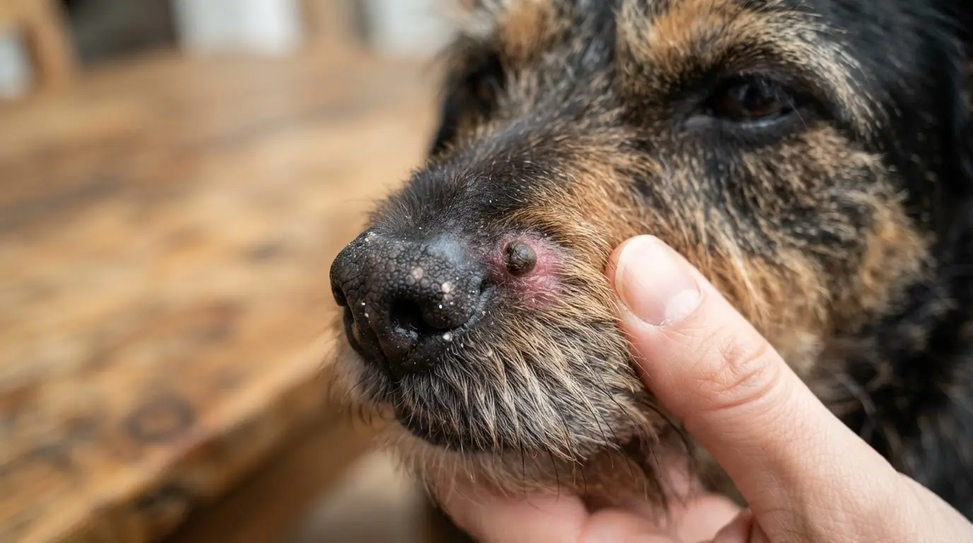
Melanomas arise from pigment-producing cells and often appear as black, brown, or pink masses on the skin, mouth, or toes.
Oral melanomas are particularly aggressive and may spread quickly. Prompt veterinary diagnosis significantly improves treatment planning and long-term outcomes.
16. Squamous Cell Carcinoma
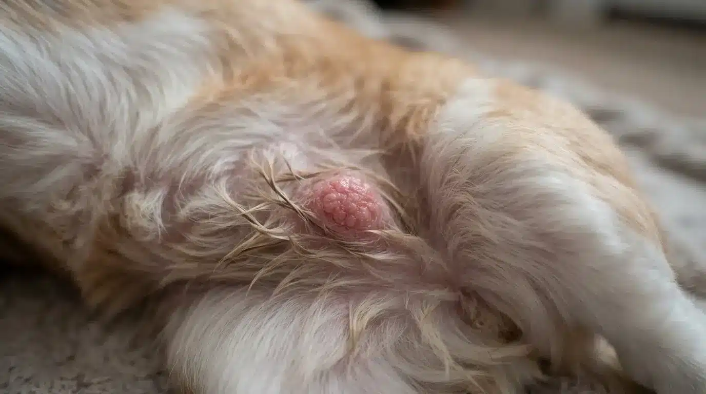
Squamous cell carcinoma usually affects lightly pigmented skin exposed to sunlight. It often appears as a crusty sore, ulcer, or raised lesion that fails to heal.
Early treatment helps prevent deeper tissue invasion and improves the chances of successful recovery.
17. Hemangiosarcoma (Skin Form)
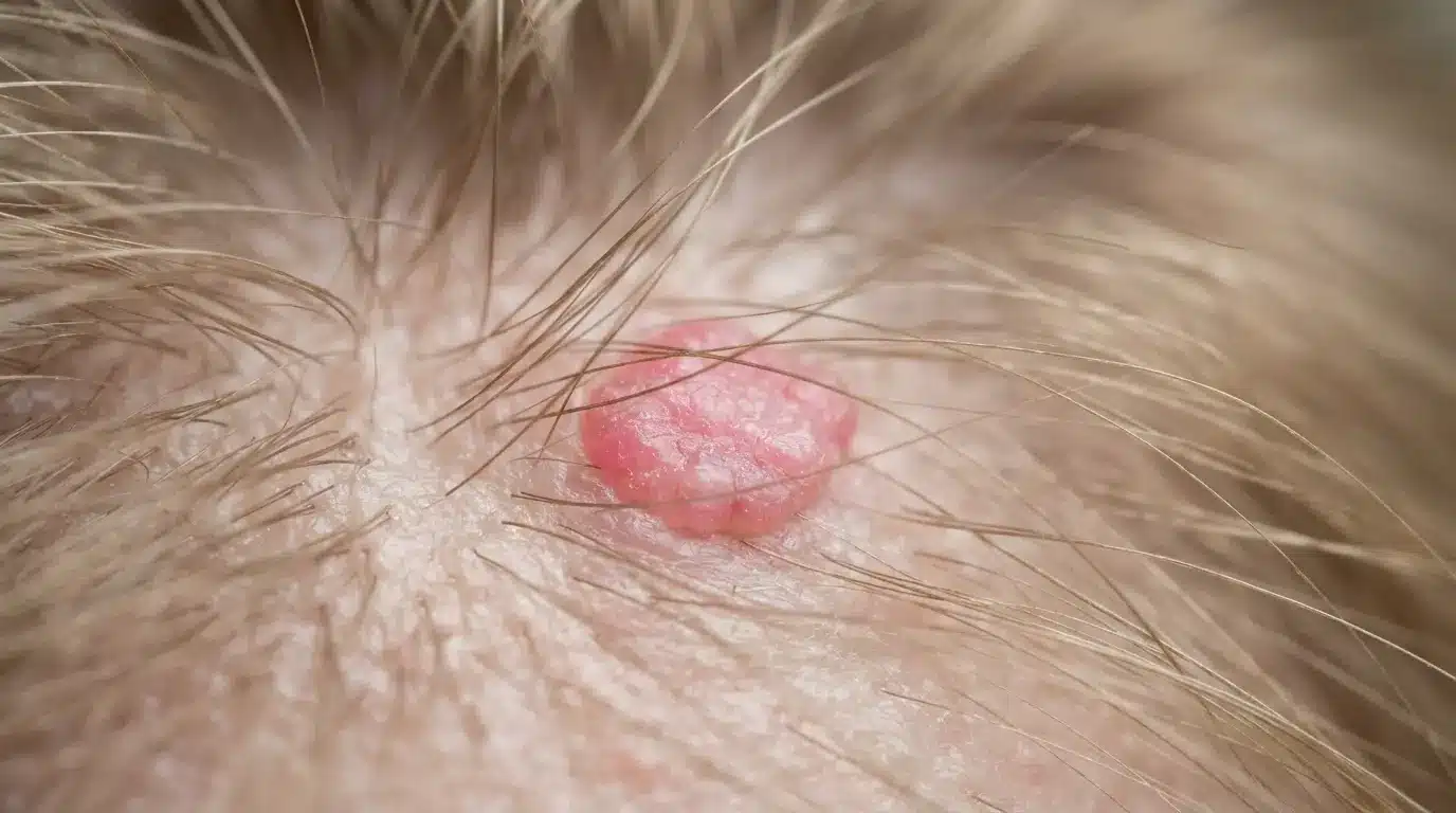
Cutaneous hemangiosarcoma develops from blood vessel cells and commonly appears as red, purple, or dark-colored skin masses.
Sun-exposed areas are more frequently affected, particularly in lightly pigmented dogs. Early surgical removal offers the best opportunity to prevent progression and improve prognosis.
18. Basal Cell Tumor
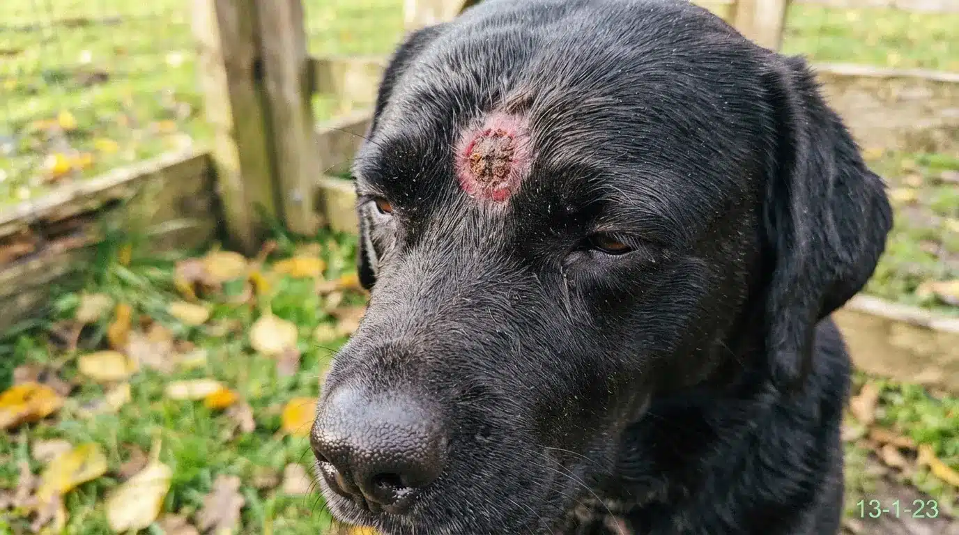
Basal cell tumors originate in the deepest layer of the skin and are usually round, raised, well-defined growths. Most are benign, although some can become locally invasive.
Veterinary examination helps determine whether monitoring or surgical removal is the most appropriate treatment.
19. Abscess
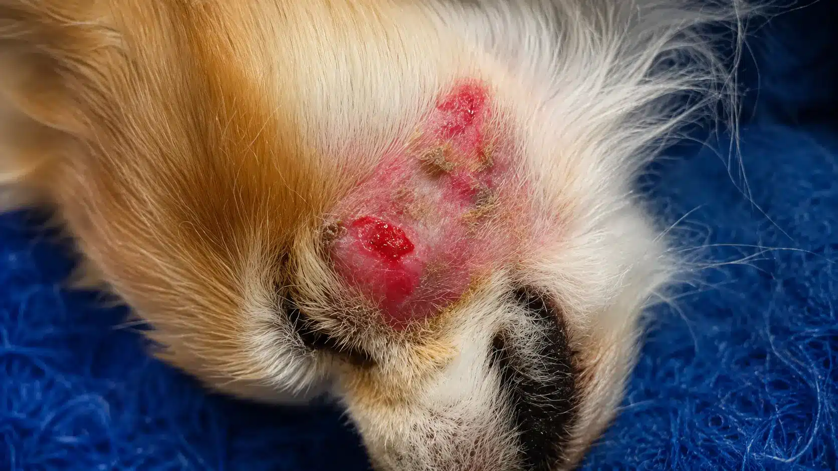
Abscesses develop when bacteria enter the skin through bites, scratches, or wounds, leading to a pocket of pus.
They usually appear as warm, swollen, painful lumps and may eventually rupture. Prompt veterinary treatment helps drain the infection and prevents further complications.
20. Skin Tag
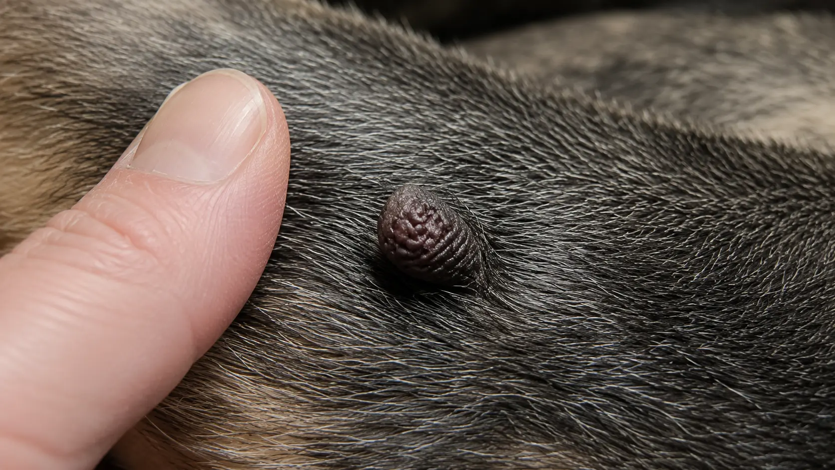
Skin tags are small, soft flaps of skin that commonly develop on older dogs. They often appear around the neck, chest, armpits, or legs and rarely cause discomfort.
Removal is usually unnecessary unless the skin tag becomes irritated, injured, or repeatedly caught during grooming.
21. Hematoma
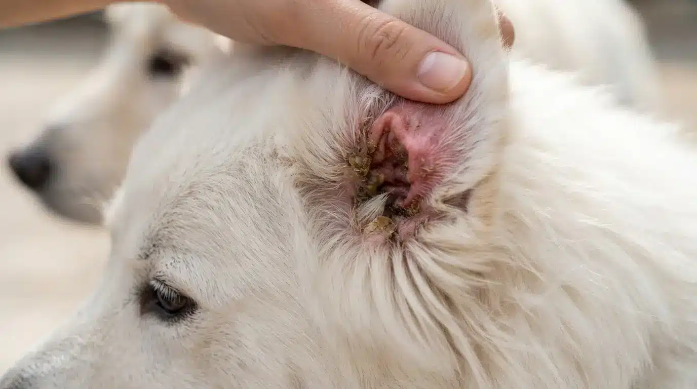
Hematomas form when blood collects beneath the skin after trauma or excessive head shaking. They appear as soft, swollen, fluid-filled lumps, especially on the ears.
Veterinary treatment may involve draining the blood and addressing the underlying cause to prevent recurrence.
Dog Cyst vs Tumor: How to Tell the Difference
Dog cysts and tumors can look very similar, making it difficult to tell them apart by appearance alone. Comparing their common characteristics provides helpful context, but only a veterinarian can confirm the exact diagnosis.
| Feature | Dog Cyst | Dog Tumor |
|---|---|---|
| What It Is | A fluid- or oil-filled sac beneath the skin. | An abnormal cell growth that may be benign or cancerous. |
| Cause | Usually caused by blocked glands or hair follicles. | Develops from abnormal cell growth, often influenced by age or genetics. |
| Appearance & Texture | Typically round, smooth, and soft to firm. | May be smooth or irregular, with a soft, firm, movable, or fixed texture. |
| Growth | Usually slow-growing and remains localized. | Can grow slowly or rapidly, depending on the type. |
| Pain & Spread | Usually painless unless infected or ruptured, and does not spread. | May become painful over time, and malignant tumors can spread to other parts of the body. |
| Diagnosis & Treatment | Diagnosed through examination or aspiration and often monitored or surgically removed. | Usually requires aspiration or biopsy, with treatment ranging from surgery to chemotherapy or radiation. |
Tips for Monitoring Dog Lumps at Home
Keeping track of changes makes veterinary visits more productive and helps identify problems early. Simple habits can provide useful details without replacing professional medical care.
- Photograph the lump under the same lighting each week.
- Measure the size with a ruler rather than estimating.
- Record the date whenever changes occur.
- Check nearby skin for additional lumps.
- Observe whether the lump feels warmer than the surrounding skin.
- Monitor your dog’s appetite, activity level, and behavior.
- Schedule regular wellness exams for senior dogs.
Small observations collected over time often reveal patterns that are difficult to remember later. Consistent monitoring supports earlier diagnosis while reducing unnecessary worry about normal day-to-day variations.
Common Mistakes When Checking Dog Lumps
Worry can lead to handling that changes the lump or delays proper testing. Avoid these common mistakes while waiting for veterinary advice or an appointment.
- Squeezing or piercing it: This may cause pain, bleeding, infection, or deeper tissue damage.
- Using human treatments: Wart removers, acne products, oils, and antiseptics may burn or poison dogs.
- Calling every soft lump a lipoma: Cancerous masses can also feel soft, smooth, or movable.
- Watching without measuring: Small changes are missed without dated photographs and consistent measurements.
- Ignoring an old lump: A familiar mass needs reassessment when its size, color, surface, or texture changes.
- Waiting for obvious illness: Dogs may act normal while a skin tumor remains small and easier to treat.
- Missing follow-up appointments: Even benign lumps that have been diagnosed should be rechecked if they change in size, color, or texture, or begin causing discomfort.
Gentle observation is enough at home. Leave draining, cutting, medicating, and firm diagnosis to the veterinary team so the growth remains safer and easier to assess.
When to Seek Veterinary Care
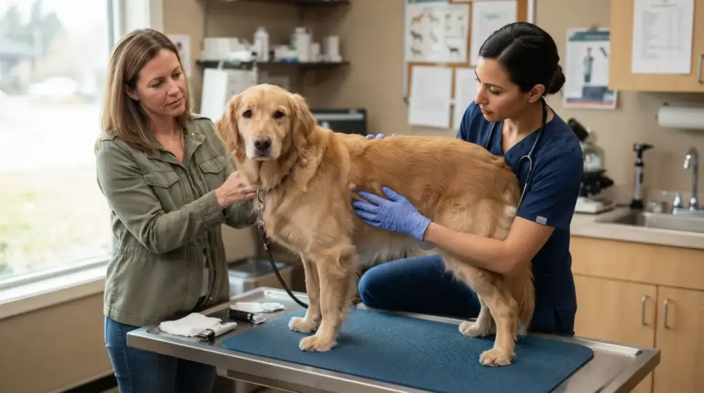
Not every lump is an emergency, but prompt veterinary care is important if a growth appears suddenly, grows quickly, changes in color or shape, becomes painful, bleeds, develops discharge, or forms an open sore.
A lump that interferes with walking, eating, or normal movement should also be examined without delay. Even if a bump looks harmless, it is worth having it checked if it persists for more than a few weeks or continues to grow.
Since many benign and cancerous growths can look alike, only a veterinarian can determine the cause through a physical examination and diagnostic tests such as fine needle aspiration or biopsy.
Early evaluation often leads to simpler treatment and better outcomes for your dog.
Final Thoughts
Watching a lump appear on your dog can feel overwhelming, but having the right information helps you respond with confidence instead of guesswork.
I hope these pictures of dog tumors and cysts have made it easier for you to compare common skin growths, understand how cysts, tumors, lumps, and bumps differ, recognize warning signs, and know when veterinary care is needed.
Remember, photos are helpful for reference, but they cannot replace a professional diagnosis. Keeping an eye on changes and acting early can make a real difference to your dog’s health.
If you found this information useful, share your thoughts in the comments and check out other pet health blogs for more helpful tips.
Frequently Asked Questions
Can Stress Cause Lumps or Cysts to Develop in Dogs?
No,stress does not directly cause lumps or cysts in dogs. However, chronic stress may weaken the immune system or worsen skin irritation, making existing skin conditions more noticeable. Any new lump should still be evaluated by a veterinarian.
Are Dog Tumors and Cysts Contagious to Other Pets?
Most tumors and cysts are not contagious and cannot spread between pets. An exception is viral papillomas (dog warts), which can spread through direct contact with infected dogs, especially in puppies or dogs with weaker immune systems.
Can Grooming Help Detect Dog Tumors and Cysts Early?
Yes. Regular grooming and brushing make it easier to notice new lumps, bumps, or skin changes hidden beneath the coat. Early detection allows a veterinarian to examine suspicious growths sooner, improving the chances of successful treatment if needed.
Do Indoor Dogs Get Tumors and Cysts?
Yes. Indoor dogs can develop tumors and cysts just as outdoor dogs can. Factors such as aging, genetics, hormonal changes, and underlying health conditions play a much greater role than whether a dog spends most of its time indoors.
Can Dog Tumors or Cysts Grow Back After Removal?
Yes. Some cysts may recur if the entire cyst lining is not removed, while certain tumors can return depending on their type and whether all abnormal tissue was successfully eliminated during surgery. Regular follow-up examinations are recommended.